Follistatin 315 vs 344: Isoforms and Heparin Binding
Follistatin isoforms differ at the C-terminus, which changes heparan sulfate binding and where the protein stays. What that means in the laboratory.


Cover illustration: AI-generated editorial artwork. Conceptual only; not an exact molecular structure, actual laboratory result or evidence of clinical effects.
The numbers in these names are residue counts, and the difference between them is a short acidic tail. That sounds trivial and is not: the tail decides whether the protein sticks to cell surfaces or circulates, which changes where it acts without changing what it binds.
For laboratory research use only. The research materials supplied by HealthyWelness are not approved medicines, foods or cosmetics and are not for human or veterinary use. This article summarises published research and is not medical advice.
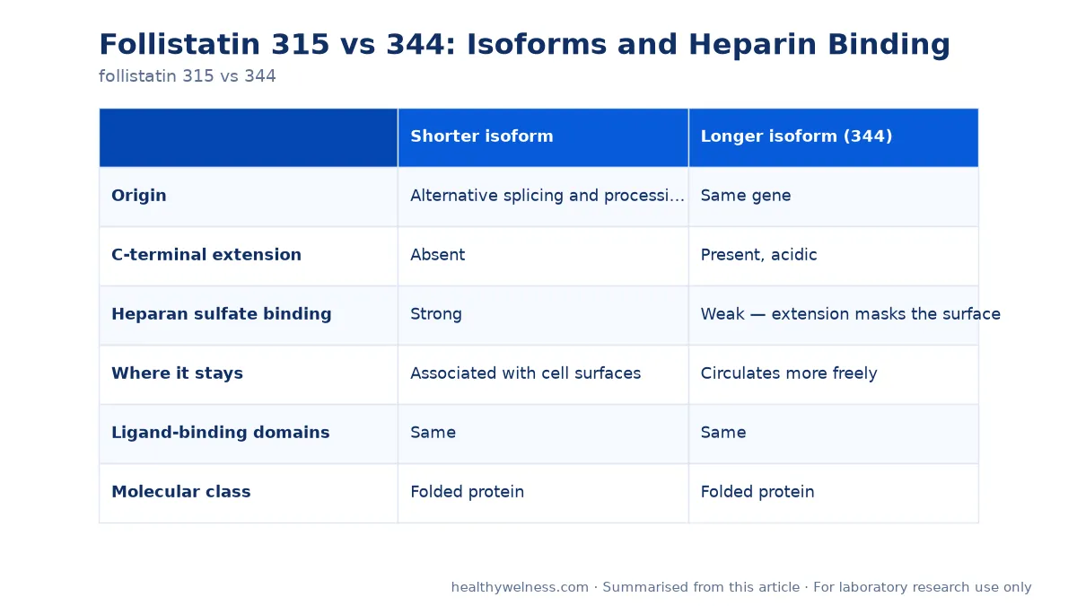
Side by side
| Shorter isoform | Longer isoform (344) | |
|---|---|---|
| Origin | Alternative splicing and processing of one gene | Same gene |
| C-terminal extension | Absent | Present, acidic |
| Heparan sulfate binding | Strong | Weak — extension masks the surface |
| Where it stays | Associated with cell surfaces | Circulates more freely |
| Ligand-binding domains | Same | Same |
| Molecular class | Folded protein | Folded protein |
What the structural work established
The 2007 study by Lerch and colleagues in the Journal of Biological Chemistry is the reference that makes this comparison interpretable. Its title states the finding directly: structural and biophysical coupling of heparin and activin binding to follistatin isoform functions.
The word doing the work is coupling. Heparin binding and ligand binding are not independent properties that happen to differ between isoforms; they are linked through the same region of the molecule. The acidic C-terminal extension of the longer isoform folds back over the basic surface that would otherwise engage heparan sulfate, so the extension acts as an intramolecular competitor.
Two consequences follow for anyone reading this literature:
- Isoform identity is a localisation variable, not an affinity variable. Both forms sequester the same ligands; they differ in where they are when they do it.
- A cell-based assay can show an isoform difference that a cell-free binding assay will not. If the readout has no cell surface and no heparan sulfate, the structural difference has nothing to act on.
Why that matters for the pathway
Follistatin binds several TGF-beta superfamily ligands, including activins and myostatin, and prevents them reaching their receptors. The 2001 PNAS paper by Lee and colleagues is the standard reference for how myostatin activity is regulated extracellularly, and the 2022 Endocrine Reviews article by Rodgers and colleagues covers the wider class of pathway-attenuating agents.
A sequestering protein held at the cell surface and one circulating in the medium produce different local ligand concentrations. In a tissue, that is the difference between acting on the cell that secreted the protein and acting at a distance. In a dish, it is the difference between an effect that washes out and one that does not.
The breadth of ligand binding remains a limitation for both isoforms. Neither is myostatin-selective, so an effect observed after adding follistatin cannot be attributed to myostatin blockade alone without controls — the point that motivated work such as the 2020 study by Saitoh and colleagues deriving a short peptide from follistatin in search of narrower selectivity. The wider comparison is in myostatin pathway research compounds.
Reading the numbers on a certificate
Isoform names in this family are a known source of confusion, because different numbering conventions circulate: some count the precursor including the signal sequence, some count the mature protein. The practical rule is the same one that applies to any fragment or variant.
| Check | Why |
|---|---|
| Stated residue count | The name alone does not fix which form is in the vial |
| Observed mass against the expected value | The isoforms differ by a measurable amount |
| Whether the numbering includes the signal sequence | Changes the number without changing the molecule |
| Expression system | Glycosylation differs, and so does the observed mass |
That last row is worth expanding. A recombinant protein expressed in a mammalian system may be glycosylated; the same sequence from a bacterial system is not. The observed mass then differs from the calculated polypeptide mass, and a mismatch is not necessarily an error. See how to read a certificate of analysis.
Handling
Both isoforms are folded proteins, not peptides, and the handling difference is categorical rather than a matter of degree:
- Do not vortex. Shear and foaming unfold proteins at the air–liquid interface, and unfolded protein aggregates irreversibly.
- Aliquot on first reconstitution. Freeze–thaw tolerance is low compared with a short peptide.
- Use carrier protein for dilute working solutions, where the assay tolerates it; proteins are lost to tube and pipette surfaces at low concentration.
- Use the diluent named on the certificate rather than a general rule.
- Expect a silent failure mode. Denatured protein has the same mass and no binding activity, so an inactive result can be a handling artefact. SDS-PAGE on a new lot is worth the hour.
Concentration maths uses the molecular weight of the exact isoform supplied, which our reconstitution calculator takes as an explicit input. Our Follistatin 344 research material is supplied for laboratory research use. Certificates are available for listed products — contact us for the latest report.
Scope note
This material is supplied for laboratory research use only. It is not a medicine, not a supplement, and not for human or veterinary use. The work discussed above is structural, biochemical and animal research.
Open questions
- How much does isoform choice change measured potency in a cell-based assay with intact heparan sulfate?
- Can the two forms be distinguished analytically in a supplied preparation?
- Does glycosylation state affect ligand sequestration as well as mass?
Related research
Frequently asked questions
What is the difference between follistatin 315 and 344?
Length, and what that length does. The numbers are residue counts of two isoforms produced from one gene by alternative splicing and processing. The longer form carries an acidic C-terminal extension that the shorter one lacks, and that extension changes how the protein interacts with cell-surface heparan sulfate.
Which isoform binds heparan sulfate more strongly?
The shorter form. Published structural and biophysical work shows the C-terminal extension of the longer isoform masks the heparin-binding surface, so the short form associates with cell surfaces and the long form circulates more freely.
Does the isoform change activin binding?
Ligand binding itself is a property both isoforms share, since the binding domains are the same. What differs is location: a cell-surface-associated protein and a freely circulating one act in different places even at the same concentration.
Why is Follistatin 344 the common research form?
It is the designation most often listed and corresponds to the precursor numbering used in much of the literature. The important point for an experiment is to read the stated residue count on the certificate rather than to assume which form a catalogue name refers to.
Are these proteins or peptides?
Proteins. Both isoforms are far larger than the short peptides in a research catalogue, and they fold. They need protein handling: gentle reconstitution, carrier protein when dilute, and as few freeze-thaw rounds as possible.
For laboratory research use only. The research materials supplied by HealthyWelness are not approved medicines, foods or cosmetics and are not for human or veterinary use. This article summarises published research and is not medical advice.
Sources
- Lerch TF, et al. Structural and biophysical coupling of heparin and activin binding to follistatin isoform functions. J Biol Chem. 2007. PMID: 17409095
- Lee SJ, et al. Regulation of myostatin activity and muscle growth. Proc Natl Acad Sci U S A. 2001. PMID: 11459935
- Rodgers BD, et al. Myostatin/Activin Receptor Ligands in Muscle and the Development Status of Attenuating Drugs. Endocr Rev. 2022. PMID: 34520530
- Saitoh M, et al. Discovery of a follistatin-derived myostatin inhibitory peptide. Bioorg Med Chem Lett. 2020. PMID: 31874826