NAD+ to NADH: Reduction or Oxidation?
NAD+ to NADH is a reduction. Why the plus sign is not a charge on the whole molecule, and why the ratio matters more than either concentration.

Cover illustration: AI-generated editorial artwork. Conceptual only; not an exact molecular structure, actual laboratory result or evidence of clinical effects.
The short answer is that NAD+ to NADH is a reduction. The longer answer explains the two things that make this question confusing in the first place: the notation, and the fact that the quantity which matters biologically is neither form on its own.
For laboratory research use only. The research materials supplied by HealthyWelness are not approved medicines, foods or cosmetics and are not for human or veterinary use. This article summarises published research and is not medical advice.
The reaction, written out
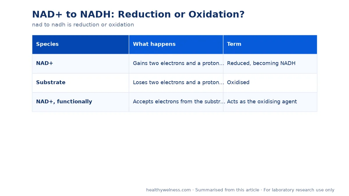
NAD+ accepts a hydride — a proton with two electrons, written H−. Formally:
| Species | What happens | Term |
|---|---|---|
| NAD+ | Gains two electrons and a proton equivalent | Reduced, becoming NADH |
| Substrate | Loses two electrons and a proton equivalent | Oxidised |
| NAD+, functionally | Accepts electrons from the substrate | Acts as the oxidising agent |
Both statements in that table are true simultaneously, and holding them together is the whole trick. NAD+ is reduced; NAD+ is the oxidising agent. These sound contradictory but describe different things — what happens to the molecule, and what role it plays for its partner. A reagent that oxidises something else is necessarily reduced in the process.
The usual mnemonic applies: reduction is gain of electrons. If it helps, note that “reduction” refers to a reduction in oxidation state, which is a number that falls as electrons are gained.
Why the notation is misleading
The plus sign in NAD+ trips up more people than the chemistry does, and the explanation is specific.
The nicotinamide ring in the oxidised form contains a nitrogen bearing a formal positive charge. That is what the plus sign records. It is a charge on one atom in one ring, not the net charge of the molecule.
The whole molecule contains two phosphate groups, which are deprotonated and negatively charged at physiological pH. So:
- NAD+ — ring nitrogen positive, phosphates negative, molecule negatively charged overall.
- NADH — ring neutral after accepting the hydride, phosphates negative, molecule negatively charged overall, and more so than NAD+.
So NADH is written without a sign because its ring is neutral, not because the molecule is. A good deal of confusion about NAD nomenclature in product descriptions traces back to reading the superscript as a net charge. The related question of NAD versus “NAD+” as product names is covered separately in NAD compared with NAD+.
Why the ratio is the quantity of interest
This is the point that matters most for interpreting the literature. NAD+ and NADH form a redox couple, and the thermodynamic driving force for any dehydrogenase reaction depends on the ratio of the two, not on the concentration of either.
The 2015 review by Bilan and colleagues covers the main cellular redox couples and is the right conceptual starting point: NAD+/NADH is one of several such couples, each maintained at its own characteristic ratio and each reporting on a different aspect of cellular state.
The consequence for experimental design is direct. Measuring NAD+ alone, or NADH alone, does not characterise redox state. A doubled NAD+ concentration with a doubled NADH concentration leaves the ratio unchanged and the thermodynamics unchanged. This is why careful studies report the ratio.
The 2017 study by Kim and colleagues in Schizophrenia Bulletin is an example of what measuring the ratio in vivo makes possible: it reported redox dysregulation detected through in vivo NAD+/NADH measurement. The 2019 review by Berthiaume and colleagues in Antioxidants and Redox Signaling takes the same approach to mitochondrial NAD+/NADH state in a cardiac context.
Compartments complicate the ratio further
There is not one NAD+/NADH ratio in a cell. The cytosolic and mitochondrial pools are separate, maintained at different ratios, and connected only by shuttle systems rather than by free diffusion of the cofactor.
The 2018 study by Goodman and colleagues in the Journal of Biological Chemistry addressed this directly, examining spatiotemporal compartmentalisation of hepatic NADH and NADPH metabolism. Two findings from that line of work matter for anyone designing a measurement:
- A whole-cell lysate measurement averages distinct compartments, and the average may not correspond to the state of either.
- NADH and NADPH are separately regulated. NADP carries an extra phosphate, is handled by different enzymes, and is maintained at a very different ratio — typically far more reduced than the NAD pool. Treating them together is a category error.
What this means for working with NAD+ as a reagent
| Consideration | Practical note |
|---|---|
| Which form is supplied | The oxidised form, NAD+, is the usual catalogue item |
| Molecular weight differs | NAD+ and NADH differ; salt forms differ again |
| Solution stability | Both forms degrade in solution; the reduced form is base-sensitive and the oxidised form acid-sensitive |
| Assay interference | NADH absorbs at 340 nm; NAD+ does not. This is the basis of most dehydrogenase assays |
| Storage | Dry, dark, frozen; prepare solutions close to use |
The absorbance difference at 340 nm is worth knowing as more than trivia: it is why the interconversion can be followed spectrophotometrically at all, and it means a preparation’s redox state can be assessed by a simple absorbance reading.
Concentration maths should use the molecular weight of the exact salt form supplied. Our reconstitution calculator takes molecular weight as an explicit input for this reason.
Our NAD+ research material is supplied for laboratory research use. Certificates are available for listed products — contact us for the latest report.
Open questions
- How closely do compartment-resolved ratios track whole-cell measurements across tissues?
- What is the most reliable method for measuring the ratio without perturbing it during sample preparation?
- How quickly does the ratio in a prepared solution shift under bench conditions?
Related research
Frequently asked questions
Is NAD+ to NADH a reduction or an oxidation?
A reduction. NAD+ gains a hydride, which means it gains two electrons and one proton equivalent, and gaining electrons is the definition of reduction. NAD+ acts as the oxidising agent in that reaction, oxidising whatever donated the hydride.
Why is the plus sign written on NAD+ but not on NADH?
Because the plus sign refers to the positive charge on the nicotinamide ring nitrogen in the oxidised form, not to the net charge of the whole molecule. The molecule carries phosphate groups and is negatively charged overall in both forms.
Does NADH have a negative charge where NAD+ has a positive one?
No. On reduction the ring nitrogen loses its formal positive charge and the ring becomes neutral. NADH is written without a sign because the ring is neutral, not because the molecule is.
Why is the NAD+/NADH ratio more informative than either concentration?
Because the pair functions as a redox couple. What determines the direction of a dehydrogenase reaction is the ratio of oxidised to reduced forms, not the absolute amount of either. Published work measures the ratio for this reason.
Are NAD and NADP the same thing?
No. NADP carries an additional phosphate and is handled by a different set of enzymes, with its own ratio maintained independently. Compartment-resolved work shows NADH and NADPH metabolism are spatially and functionally distinct.
References
- Berthiaume JM, et al. Mitochondrial NAD(+)/NADH Redox State and Diabetic Cardiomyopathy. Antioxid Redox Signal. 2019. PMID: 29073779 https://pubmed.ncbi.nlm.nih.gov/29073779/
- Goodman RP, et al. Spatiotemporal compartmentalization of hepatic NADH and NADPH metabolism. J Biol Chem. 2018. PMID: 29514978 https://pubmed.ncbi.nlm.nih.gov/29514978/
- Bilan DS, et al. Main Cellular Redox Couples. Bioorg Khim. 2015. PMID: 26615634 https://pubmed.ncbi.nlm.nih.gov/26615634/
- Kim SY, et al. Redox Dysregulation in Schizophrenia Revealed by in vivo NAD+/NADH Measurement. Schizophr Bull. 2017. PMID: 27665001 https://pubmed.ncbi.nlm.nih.gov/27665001/
For laboratory research use only. The research materials supplied by HealthyWelness are not approved medicines, foods or cosmetics and are not for human or veterinary use. This article summarises published research and is not medical advice.
Sources
- Berthiaume JM, et al. Mitochondrial NAD(+)/NADH Redox State and Diabetic Cardiomyopathy. Antioxid Redox Signal. 2019. PMID: 29073779
- Goodman RP, et al. Spatiotemporal compartmentalization of hepatic NADH and NADPH metabolism. J Biol Chem. 2018. PMID: 29514978
- Bilan DS, et al. Main Cellular Redox Couples. Bioorg Khim. 2015. PMID: 26615634
- Kim SY, et al. Redox Dysregulation in Schizophrenia Revealed by in vivo NAD+/NADH Measurement. Schizophr Bull. 2017. PMID: 27665001