Thymosin Alpha-1 Reconstitution and Concentration Guide
How to reconstitute thymosin alpha-1 for laboratory work: diluent choice, concentration arithmetic, aliquoting, and what stability studies show.

Cover illustration: AI-generated editorial artwork. Conceptual only; not an exact molecular structure, actual laboratory result or evidence of clinical effects.
Reconstituting thymosin alpha-1 means adding a measured diluent volume to the lyophilised vial and calculating the resulting solution concentration from the net peptide content on the certificate, not from the label strength. The peptide is a defined synthetic sequence, so the arithmetic is straightforward; the choices that matter are diluent, aliquoting and storage.
For laboratory research use only. The research materials supplied by HealthyWelness are not approved medicines, foods or cosmetics and are not for human or veterinary use. This article summarises published research and is not medical advice.
- Thymosin alpha-1 is a synthetic polypeptide with a reported serum half-life of approximately two hours (Ancell et al., 2001).
- Solution concentration equals net peptide content divided by diluent volume: 5 mg in 2 mL gives 2.5 mg/mL.
- Thymosin alpha-1 microparticles in PLGA implants showed 29.3% release in the first 24 hours and continuous release to 28 days in rats, with an in vitro to in vivo correlation coefficient of 0.9899 (Liu et al., 2010).
- The peptide occurs naturally in the thymus and is produced in vivo by cleavage of prothymosin alpha (Dominari et al., 2020).
- Camerini and Garaci describe the safety profile of thymosin alpha-1 as excellent and virtually devoid of toxicity across the clinical literature (Camerini and Garaci, 2015).
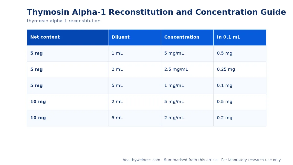
Concentration arithmetic
| Net content | Diluent | Concentration | In 0.1 mL |
|---|---|---|---|
| 5 mg | 1 mL | 5 mg/mL | 0.5 mg |
| 5 mg | 2 mL | 2.5 mg/mL | 0.25 mg |
| 5 mg | 5 mL | 1 mg/mL | 0.1 mg |
| 10 mg | 2 mL | 5 mg/mL | 0.5 mg |
| 10 mg | 5 mL | 2 mg/mL | 0.2 mg |
Every figure in that table is simple division, and the peptide calculator does the same arithmetic for any vial and target concentration. The error that matters is not in the division but in the numerator: using the label strength rather than the net peptide content on the certificate overstates how much peptide is present, because the gross figure includes counterion mass and residual water. For a trifluoroacetate salt that difference can be substantial, as explained in peptide counterions: TFA vs acetate.
Material from the thymosin alpha-1 research peptide listing is supplied as a lyophilised powder for laboratory use only. Certificates are available for listed products — contact us for the latest report.
Procedure
| Step | Action | Reason |
|---|---|---|
| 1 | Equilibrate vial to room temp | Avoids condensation |
| 2 | Read net content on certificate | Sets the numerator |
| 3 | Calculate diluent volume | Fixes concentration |
| 4 | Add diluent down the wall | Limits foaming |
| 5 | Swirl, do not shake | Shear damages peptides |
| 6 | Inspect against light | Haze signals a problem |
| 7 | Aliquot and label | Limits repeated warming |
Two of those steps deserve expansion. Adding the diluent against the vial wall rather than directly onto the cake slows the wetting front and reduces the air-liquid interface where surface denaturation occurs; it costs nothing and removes a common cause of cloudy solutions. And inspection is a genuine test rather than a formality: a correctly reconstituted peptide solution at these concentrations is clear and colourless, so visible haze, fibres or particles mean the material should be set aside rather than used.
Labelling should record the net content used, the diluent, the volume, the resulting concentration and the date. Without those five items an aliquot found in a freezer three months later is unusable, since its concentration cannot be reconstructed. General practice is in how to reconstitute peptides and reconstituted peptide storage.
Choosing the diluent
The three realistic options serve different purposes. Sterile water is the neutral choice when the solution will be used in the same session and nothing in the assay objects to an unbuffered diluent. Bacteriostatic water containing 0.9% benzyl alcohol is the choice when a working solution needs to survive repeated sampling over days, because the preservative limits microbial growth in a vial that is entered more than once. Assay buffer is correct whenever the experiment specifies a pH or ionic strength, and in that case the buffer, not the peptide, dictates the choice.
Benzyl alcohol is the variable to watch. It is useful in a multi-day vial and unhelpful in a cell-based assay, where it is an additional compound in the medium at a concentration that depends on how much solution was added. If the readout is cellular, an unpreserved diluent avoids a confound that is otherwise easy to overlook. The comparison is set out in bacteriostatic water vs sterile water, and the preservative’s own properties in what is bacteriostatic water.
What the formulation literature implies for handling
Liu and colleagues’ PLGA implant study was not designed as a stability guide, but its findings are informative for bench work. The authors prepared thymosin alpha-1 microparticles by spray-freeze drying with chitosan or bovine serum albumin as a protectant before suspending them in PLGA solutions — meaning a protectant was considered necessary to carry the peptide through processing. The optimal formulation gave 29.3% release in the first 24 hours and then slow continuous release to 28 days in Sprague-Dawley rats, with in vitro and in vivo profiles correlating at 0.9899.
Three practical readings follow. The peptide tolerates processing when a protein or polysaccharide protectant is present, which supports the common practice of including carrier protein in very dilute working solutions. Its own circulating persistence is short, about two hours by serum half-life, which is why sustained-release work exists at all and why a published study’s sampling schedule is tied to its formulation. And a peptide that can be held stably inside a polymer matrix for 28 days is not therefore stable for 28 days in aqueous solution at room temperature — the matrix is doing the work.
Dilution for low-concentration assays
Where an assay needs micromolar or nanomolar concentrations, serial dilution from a stock is more accurate than attempting to weigh or reconstitute a very small amount. Two points keep those dilutions honest. Adsorption to plastic becomes significant at low peptide concentrations, which is the main reason carrier protein or a low concentration of a non-interfering surfactant is added to dilution buffers. And each dilution step carries its own pipetting error, so fewer, larger steps are usually more accurate than many small ones.
A worked route: reconstitute to a known stock in mg/mL, convert to molar concentration using the peptide’s formula mass for the salt form supplied, then dilute in the assay buffer immediately before use. The conversion step is where the salt form matters again, since the same mass of two salt forms contains different amounts of peptide. Wider context on the compound and its research record is in what is thymosin alpha-1 and the thymosin alpha-1 research review, and the comparison with thymus extracts in thymalin vs thymosin alpha-1.
Recording what was done
A reconstitution record is part of the experiment, not paperwork around it. Five entries make a solution reusable by someone else or by the same person weeks later: the net peptide content taken from the certificate, the diluent and whether it was preserved, the volume added, the calculated concentration, and the date of preparation. A sixth entry is worth adding where relevant — the lot identifier on the vial — because without it a later question about a specific result cannot be traced back to the material that produced it.
Two conventions prevent most downstream confusion. Record concentration in both mass and molar terms, since protocols are written either way and converting later requires the formula mass for the correct salt form. And note the appearance of the solution at the time of preparation, because a later observation of haze means something different depending on whether the solution was clear to begin with.
Frequently asked questions
How is solution concentration calculated for a thymosin alpha-1 vial?
Divide the net peptide content stated on the certificate by the diluent volume added. A vial with 5 mg net content reconstituted in 2 mL gives 2.5 mg/mL, and 0.1 mL of that solution contains 0.25 mg. The label strength is not the figure to use, because salt and residual moisture are included in it.
Which diluent should be used?
That depends on the protocol, not on the peptide. Sterile water suits same-session use; bacteriostatic water containing 0.9% benzyl alcohol is used where a multi-day working solution is intended. Assay buffer is appropriate when the experiment specifies it, and benzyl alcohol should be avoided where it would interfere with a cell-based readout.
Does thymosin alpha-1 need to be aliquoted?
Aliquoting is good practice for any peptide stock that will be sampled repeatedly, because each warming and cooling of the whole vial exposes all of the material to the same stress. Single-use aliquots limit that exposure to the portion being used.
How long does thymosin alpha-1 persist once administered in published studies?
Reported serum half-life is about two hours, with peak concentrations within two hours of administration and a return to baseline within 24 hours (Ancell et al., 2001). That short persistence is why sustained-release formulations have been investigated.
What do the formulation studies suggest about stability?
Liu and colleagues developed poly(lactide-co-glycolide) in situ forming implants containing thymosin alpha-1 microparticles, using chitosan or bovine serum albumin as a protectant, and reported an initial release of 29.3% over 24 hours followed by continuous release up to 28 days in rats (Liu et al., 2010).
For laboratory research use only. The research materials supplied by HealthyWelness are not approved medicines, foods or cosmetics and are not for human or veterinary use. This article summarises published research and is not medical advice.
Sources
- Ancell CD, et al. Thymosin alpha-1. Am J Health Syst Pharm. 2001. PMID: 11381492
- Liu Q, et al. In vitro and in vivo study of thymosin alpha1 biodegradable in situ forming poly(lactide-co-glycolide) implants. Int J Pharm. 2010. PMID: 20650309
- Dominari A, et al. Thymosin alpha 1: A comprehensive review of the literature. World J Virol. 2020. PMID: 33362999
- Camerini R, et al. Historical review of thymosin alpha 1 in infectious diseases. Expert Opin Biol Ther. 2015. PMID: 26098768