Buffer Capacity vs pH: Interpreting Peptide Assay Media
Learn why equal pH does not mean equal buffer capacity in peptide research, with a worked acid-load example and checks for ionic-strength confounding.

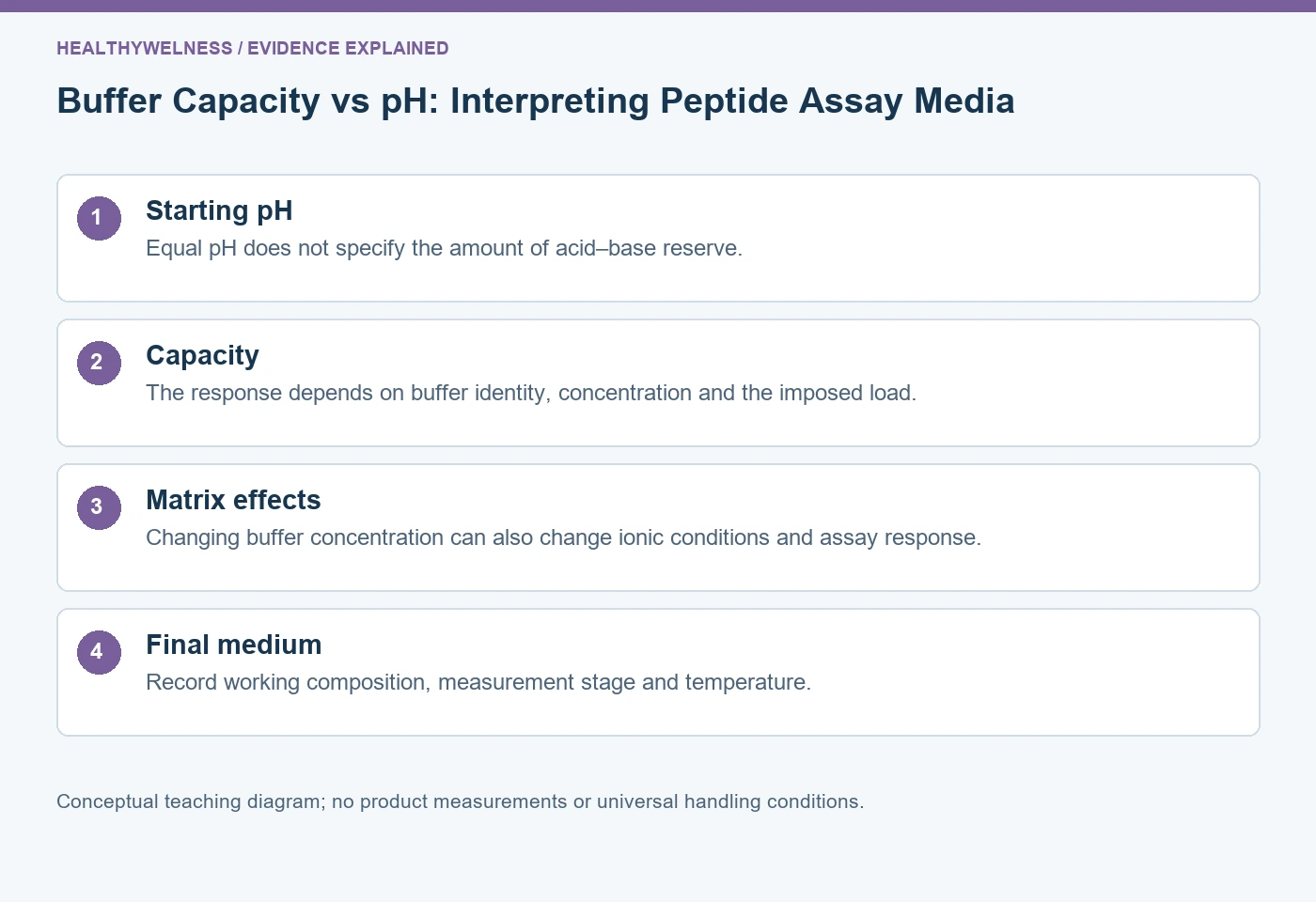
pH describes a solution’s acid–base condition; buffer capacity describes its resistance to a pH change when acid or base is added. Two peptide assay media can begin at the same pH yet respond very differently to the same acid load. Buffer identity, total concentration, temperature and other ions must be considered separately from the initial meter reading.
- Equal starting pH does not establish equal resistance to an acid or base load.
- For a simple weak-acid buffer model, pH depends on the base-to-acid ratio while reserve also depends on total concentration.
- Changing buffer concentration can change the assay matrix as well as its capacity.
- A final reaction medium may have a different pH and composition from the stock buffer used to prepare it.
The meter reading does not measure the acid–base reserve
A preparation labelled “pH 7.0” tells only part of its story. That reading does not state how much weak acid and conjugate base are present, how much acid or base the preparation can accommodate, or what will happen after all assay components are combined. It also does not identify the peptide’s isoelectric point, which concerns molecular charge balance rather than the solution’s reserve.
In the simple weak-acid model, the Henderson–Hasselbalch relation expresses pH as pKa + log10([base]/[acid]). The ratio can remain the same while both concentrations change by a factor of ten. Thus, two model buffers can have the same pH but different quantities of base available to accept an added proton load. MilliporeSigma’s Buffer Reference Center provides the acid–base relation and buffer-specific pKa and temperature information. [1]
The relation is a model with conditions, not an instruction to replace measurement with arithmetic. Activities, ionic conditions, multiple equilibria and temperature can matter in actual media. Its value here is explanatory: a ratio cannot specify the total amount represented by that ratio.
| Property | Meaning in the comparison | Useful record |
|---|---|---|
| pH | Acid–base condition at measurement | Value and temperature |
| Total buffer concentration | Amount of buffer species per volume | Final working concentration |
| Buffer capacity | Resistance to a defined perturbation | Model or measured response |
| Ionic composition | Buffer ions, salts and counterions | Complete final medium |
Capacity is a response to a perturbation
Buffer capacity is often expressed as the amount of strong acid or base per unit volume required for a unit pH change, interpreted locally around a specified condition. A finite acid-addition experiment instead measures a change over a stated interval. These descriptions are related, but a large challenge can move the system away from the starting condition, where the local capacity is different.
ITW Reagents describes the dependence of capacity on concentration and proximity to pKa. Its selection guidance also warns that increasing buffer concentration and the associated ionic environment can affect enzyme behaviour. These are first-party technical considerations, not a universal concentration recommendation for every peptide assay. [2]
For the simplified monoprotic model, acid and base are balanced at pH = pKa, giving the greatest capacity for that fixed total buffer concentration. Moving away from that point makes one member of the pair less abundant. The reserve against one direction of perturbation then becomes more limited. This is a smooth change, not an abrupt disappearance of all buffering at an arbitrary pH boundary.
The direction of the load matters. A preparation rich in conjugate base has more capacity to accommodate added acid than its scarce acid component has to accommodate added base. A single description such as “strong buffer” loses that context. State the starting condition, perturbation and acceptable pH interval for the research question.
Worked example: equal pH, unequal response to an acid load
Hypothetical ideal model: consider an invented monoprotic buffer with pKa 7.00. Ignore activity-coefficient effects, water autoionization, temperature change, volume change and all other proton-binding species. Medium A contains 10 mM total buffer, split into 5 mM acid and 5 mM base. Medium B contains 100 mM total buffer, split into 50 mM acid and 50 mM base. Both begin at calculated pH 7.00.
Now stipulate an identical 1 mM strong-acid equivalent introduced into each final medium with negligible volume change. In this model, it converts 1 mM conjugate base to acid. Medium A then contains 4 mM base and 6 mM acid. Its calculated pH is 7.00 + log10(4/6), approximately 6.824. Medium B contains 49 mM base and 51 mM acid, giving 7.00 + log10(49/51), approximately 6.983.
| Model medium | Initial total buffer | Final base / acid | Calculated final pH |
|---|---|---|---|
| A | 10 mM | 4 / 6 mM | 6.824 |
| B | 100 mM | 49 / 51 mM | 6.983 |
The calculated changes are about −0.176 and −0.017 pH units. Equal starting pH did not produce equal response to the same imposed load. The calculation illustrates a capacity difference without implying that either medium suits a real assay. The selected pKa, concentrations and acid load are teaching inputs, not measured properties of a named buffer or peptide product.
The model also does not predict a peptide’s activity, solubility or chemical stability. It contains no binding reaction, surface interaction or optical detector. If a real experiment gives different signals in A and B, the calculation supplies a plausible reason to check pH behaviour; it cannot prove that pH caused the difference.
Higher capacity can change more than the pH trajectory
Increasing total buffer concentration changes chemical composition. Depending on the buffer forms and pH adjustment, it may also change concentrations of charged species and counterions. Ionic strength depends on the concentrations and squared charges of the ions present, not simply on the pH. Two media at the same pH therefore need not provide the same electrostatic environment.
This creates a confounding problem in peptide assays. If one medium produces a larger signal and also contains more buffer, possible explanations include a different pH trajectory, direct buffer interactions, altered ionic conditions or an effect on the readout. The observation does not identify which explanation is responsible. A successful comparison records the final matrix and assesses the particular alternatives relevant to its method.
Merck’s reproducibility guidance discusses buffer identity, working concentration, temperature, metal interactions, additives and downstream detection. It recommends preserving preparation and measurement details in published methods. These considerations support checking compatibility rather than assuming a buffer is chemically invisible because its pH is correct. [3]
- A buffer-only blank addresses background from that medium in the selected readout.
- A matched vehicle comparison addresses components introduced with the peptide preparation.
- A pH observation in the final medium addresses the environment after mixing.
- An appropriate alternative readout can help assess a detection-specific explanation.
None of those comparisons proves everything. A clean blank does not exclude a buffer–analyte interaction, and unchanged pH does not demonstrate unchanged ionic composition. Choose comparisons that target the claim being made.
Measure and document the medium at the relevant stage
A stock buffer is not the final assay. Mixing it with peptide stock, cofactors, substrate, salts and other components changes the final concentrations. Some additions can also introduce acid or base equivalents. Preserve the final working composition rather than reporting only a stock label copied from a bottle.
Temperature needs the same care. A pH value obtained under one temperature does not necessarily describe the medium at another, because buffer equilibria can change. The Buffer Reference Center lists different temperature dependencies for named buffers. That supports recording temperature and checking the actual context; it does not justify one correction factor for every formulation. [1]
Measurement itself has practical limits. A small-volume assay may not accommodate a conventional electrode without altering the sample. A representative preparation can help only if it actually preserves the composition and conditions relevant to the claim. Report that relationship clearly instead of presenting an extrapolated value as a direct measurement of every assay well.
When comparing publications, missing detail should remain missing detail. If a paper says only “buffer at pH 7,” one cannot reconstruct capacity, salt composition or temperature from that phrase. The evidence may still support its reported observation, but transfer to another preparation is uncertain. Do not fill the gaps with familiar laboratory defaults.
Decide whether the question is pH control or matrix compatibility
Begin with the uncertainty. If the concern is pH movement after addition, examine the final medium’s response to the relevant perturbation. If pH remains comparable but signal changes with buffer concentration, investigate matrix and detection explanations. If changing buffer identity changes the result, preserve both its acid–base properties and its other chemistry in the interpretation.
Define a suitable pH interval from the actual research method, then assess whether the medium maintains it without introducing unacceptable interference. More capacity is useful only relative to that objective. It is not a free-standing quality score, and this article provides no universal buffer choice, concentration or storage instruction.
Questions about pH and capacity in peptide research
Can two buffers at pH 7 have different capacities?
Yes. Equal pH does not fix the total concentrations or buffer identities. Different acid–base reserves can produce different responses to the same added acid or base.
Does adding more salt necessarily improve buffering?
No. Ionic composition and acid–base buffering are distinct. A salt that does not provide the relevant acid–base pair cannot be assumed to supply the needed capacity.
Is the highest buffer concentration always preferable?
No. Increasing concentration can affect ionic conditions, interactions and detection. Suitability depends on maintaining the required conditions while preserving the validity of the assay.
Can I infer final assay pH from the stock buffer label?
Not reliably without evidence that the added components and working conditions preserve it. Report the final medium and relevant measurement stage rather than only the stock description.
Does a peptide’s pI determine buffer capacity?
No. A peptide’s pI concerns its average charge balance. Capacity concerns the solution’s response to acid or base and depends on the species and amounts present.
Read the related charge and measurement guides
Continue with peptide pI versus pH, controls in peptide experiments and fluorescence interference. Use the peptide concentration calculator for nominal peptide mass–volume arithmetic; it does not calculate buffer capacity or predict final pH. HealthyWelness operates a commercial research-material catalog. The worked example is educational reasoning and reports no product testing.
References
- MilliporeSigma. Buffer Reference Center.
- ITW Reagents. Buffers: criteria for selection and setting the pH value.
- Merck. Reproducibility with Biological Buffers.
Cover and evidence map: original conceptual editorial schematics, not measured results, exact molecular structures, or photographs of HealthyWelness facilities.