GHRP-6 and GHRP-2 Reconstitution and Mixing
Both peptides are tryptophan-rich hexapeptides with D-residues. Why that makes them light-sensitive and why mass alone cannot confirm them.

Cover illustration: AI-generated editorial artwork. Conceptual only; not an exact molecular structure, actual laboratory result or evidence of clinical effects.
GHRP-6 and GHRP-2 are both six-residue peptides from the same series, and they take the same handling because they share the same two liabilities: tryptophan, which makes them light- and oxidation-sensitive, and D-amino acids, which make mass alone insufficient to confirm identity. Everything below follows from those two facts.
For laboratory research use only. The research materials supplied by HealthyWelness are not approved medicines, foods or cosmetics and are not for human or veterinary use. This article summarises published research and is not medical advice.
- GHRP-6 is His-D-Trp-Ala-Trp-D-Phe-Lys-NH2, a six-residue peptide with two tryptophan residues and a histidine.
- Both GHRP-6 and GHRP-2 contain D-amino acids, and D-to-L substitution does not change molecular mass.
- Metal-catalysed oxidation at histidine is a documented degradation route in model peptides (Khossravi et al., 1998, PMID 9688066).
- Freeze-dried peptide stability is governed by moisture and oxygen content, not storage temperature alone (Pikal et al., 1992, PMID 1592171).
- Both peptides are usually supplied as acetate salts, so counterion mass is part of the vial weight.
How should either peptide be reconstituted?
- Let the sealed vial reach room temperature before opening. Opening cold glass in a humid room condenses water into a hygroscopic powder, and that moisture does not leave again.
- Centrifuge briefly if the cake may have moved in transit.
- Use the diluent named on the certificate, and prefer a clean one. Old buffer stocks are the commonest unnoticed source of trace metal.
- Add the diluent slowly down the inside wall, letting it run over the cake.
- Swirl until dissolved. Do not vortex a tryptophan-rich peptide harder than necessary.
- Keep it out of direct light during preparation, and return it to an amber vial or foil-wrapped tube.
- Aliquot immediately into single-use volumes and freeze; date and label each tube.
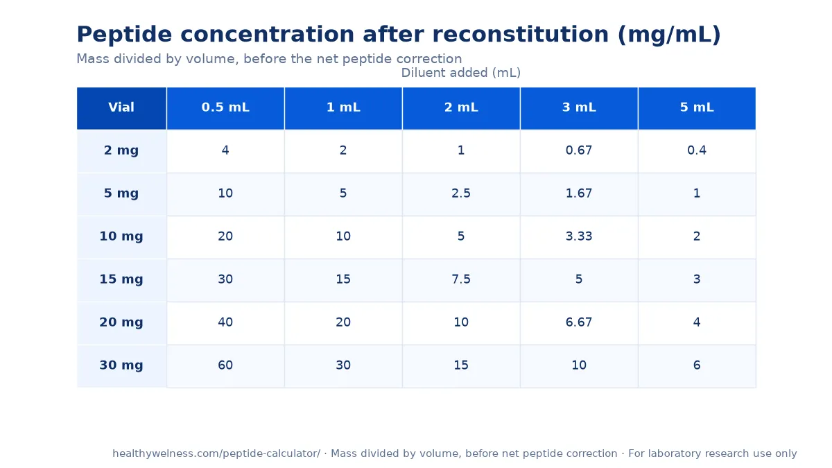
The chart above gives the gross mass-over-volume arithmetic for common vial sizes. The net peptide content correction, which is what makes the figure accurate, is applied by our reconstitution calculator.
Why does tryptophan content change the precautions?
GHRP-6 carries two tryptophan residues out of six, plus a histidine. GHRP-2 shares the same design family and the same liability. Tryptophan and histidine are the two residues most readily oxidised in peptides, and tryptophan also absorbs in the ultraviolet, which makes photo-oxidation a route as well.
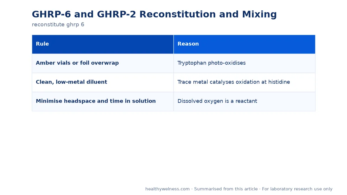
The 1998 work by Khossravi and colleagues established metal-catalysed oxidation of histidine as a defined degradation pathway in model peptides. Translated into practice, that gives three rules with reasons rather than three habits:
| Rule | Reason |
|---|---|
| Amber vials or foil overwrap | Tryptophan photo-oxidises |
| Clean, low-metal diluent | Trace metal catalyses oxidation at histidine |
| Minimise headspace and time in solution | Dissolved oxygen is a reactant |
None of this produces a visible change. An oxidised preparation looks exactly like a fresh one, which is why the discipline has to be preventive rather than reactive. For contrast, ipamorelin contains neither tryptophan nor histidine and is correspondingly forgiving — see ipamorelin stability and storage.
Why can mass spectrometry not confirm these peptides?
Because both contain D-amino acids by design, and changing the handedness of an amino acid does not change its formula. A batch synthesised with the wrong stereochemistry at a D-position has exactly the correct molecular mass and will pass a mass-only confirmation.
What resolves it is chromatographic retention: stereoisomers separate on a reverse-phase column even though they cannot be separated by mass. So for this peptide family the useful request is both numbers plus the trace:
- Observed mass as a number, compared against a value you compute for the stated sequence and terminal form.
- The chromatogram, not only a purity percentage, so a single clean peak can be seen rather than inferred.
- Chirality verification where the work depends on it, which is a separate test that is rarely performed unless asked for.
The framework is in how to read a certificate of analysis, and the molecular background in what GHRP-6 is.
How should solutions be stored?
| State | Condition | Why |
|---|---|---|
| Sealed lyophilized | −20 °C, desiccated, dark | Moisture and oxygen govern solid-state stability |
| Before each opening | Warm the sealed vial first | Prevents condensation into the powder |
| Opened lyophilized | Reseal with desiccant, return to −20 °C | Hygroscopic once exposed |
| Reconstituted, in use | Refrigerated, dark, short period | Days of working use, not months |
| Reconstituted, stored | Aliquoted, frozen, dated | One warming round per aliquot |
| Dilute working solution | Prepare fresh | Surface loss and faster degradation |
The solid-state rows rest on measurement. The 1992 study by Pikal and colleagues found freeze-dried stability governed by moisture and oxygen content rather than temperature alone, which is the argument for desiccant and a properly reseated cap rather than reliance on the freezer. For solutions, the 1998 study by Gietz and colleagues on degradation kinetics shows how strongly pH governs rate — so the buffer is a stability variable worth recording as part of the method.
Why does the counterion correction matter for a hexapeptide?
Because counterion mass is a cost per charged residue and a six-residue peptide has few residues to spread it over. The default is trifluoroacetate, left behind by reverse-phase purification unless an exchange step is added; the 2008 evaluation by Roux and colleagues found exchange approaches to differ in how completely they succeed.
So a vial labelled with a nominal weight contains the peptide plus counterion plus residual water, and the non-peptide share is proportionally larger here than for a long peptide. Using gross weight produces a solution more dilute than intended, systematically and always in the same direction. The detail is in peptide counterions: TFA versus acetate.
Our GHRP-6 research material is supplied for laboratory research use. Certificates are available for listed products — contact us for the latest report.
Why a six-residue peptide still needs discipline
Short peptides have a reputation for robustness, and in one sense it is deserved: there is no fold to lose and no disulfide to scramble. That reputation causes trouble here, because the specific liabilities of these two have nothing to do with size.
| Assumption about short peptides | True for these two? |
|---|---|
| No conformational failure mode | True |
| Tolerant of vigorous mixing | Partly — oxidation risk argues against it |
| Identity settled by mass alone | False — D-residues are mass-silent |
| Not especially light-sensitive | False — two tryptophan residues |
| Counterion is a rounding detail | False — proportionally large at this length |
Three of those five assumptions are wrong for this pair, which is why the handling list above is longer than a hexapeptide appears to warrant. None of the precautions is expensive; the cost is remembering that length and fragility are not the same axis.
A final practical note: record the diluent, the date and the concentration on every aliquot. When a result later looks wrong, that label is usually the fastest route to an explanation.
Scope note
This material is supplied for laboratory research use only. It is not a medicine, not a supplement, and not for human or veterinary use. The 1998 review by Bowers is cited as background on how this peptide series was developed.
Open questions
- How quickly does tryptophan oxidation become detectable under ordinary bench lighting?
- Is chiral verification practical as a routine release test at this peptide length?
- How much do GHRP-6 and GHRP-2 differ in measured stability under identical conditions?
Related research
Frequently asked questions
How is GHRP-6 reconstituted?
Warm the sealed vial to room temperature, add the diluent named on the certificate down the inside wall, swirl until dissolved rather than vortexing, inspect for haze, then aliquot into single-use tubes and freeze. Concentration is computed from net peptide content, not the nominal vial weight.
Do GHRP-6 and GHRP-2 need different handling?
No. Both are tryptophan-rich hexapeptides containing D-amino acids and supplied as acetate salts, so the same precautions apply: light protection, a low-metal diluent, gentle mixing and aliquoting on first use.
Why are these peptides light-sensitive?
Because of their tryptophan content. Tryptophan is among the most readily oxidised amino acids and absorbs in the ultraviolet, so a tryptophan-rich peptide is both photo-sensitive and oxidation-prone. Amber vials or foil overwrap are a real precaution, not a convention.
Why does a low-metal diluent matter?
Trace metal ions catalyse oxidation, and these peptides contain both histidine and tryptophan, the two residues most affected. Using a clean diluent and avoiding old buffer stocks removes a degradation route that produces no visible change.
Can mass spectrometry confirm that the material is correct?
Only partly. Both peptides contain D-amino acids, and changing the handedness of a residue does not change molecular mass. A batch with the wrong stereochemistry has the correct mass; chromatographic retention is what resolves it.
For laboratory research use only. The research materials supplied by HealthyWelness are not approved medicines, foods or cosmetics and are not for human or veterinary use. This article summarises published research and is not medical advice.
Sources
- Bowers CY. Growth hormone-releasing peptide (GHRP). Cell Mol Life Sci. 1998. PMID: 9893708
- Khossravi M, et al. Chemical pathways of peptide degradation: IX. Metal-catalyzed oxidation of histidine in model peptides. Pharm Res. 1998. PMID: 9688066
- Pikal MJ, et al. Formulation and stability of freeze-dried proteins: effects of moisture and oxygen on the stability of freeze-dried formulations of human growth hormone. Dev Biol Stand. 1992. PMID: 1592171
- Roux S, et al. Elimination and exchange of trifluoroacetate counter-ion from cationic peptides: a critical evaluation of different approaches. J Pept Sci. 2008. PMID: 18035848
- Gietz U, et al. Chemical degradation kinetics of recombinant hirudin (HV1) in aqueous solution: effect of pH. Pharm Res. 1998. PMID: 9755900