Glutathione and Inflammation: Thiol Signalling in Immune Cells
How glutathione enters inflammatory signalling: S-glutathionylation of IKK-beta, GSTP and GSTO1 in macrophages, and the airway models behind the claims.

Cover illustration: AI-generated editorial artwork. Conceptual only; not an exact molecular structure, actual laboratory result or evidence of clinical effects.
Glutathione takes part in inflammation as a signalling molecule, not only as an antioxidant: it is attached to and removed from reactive cysteines on proteins such as IKK-beta, which changes how NF-kappaB pathways respond. The evidence comes mainly from cell lines, macrophages and mouse airway models, and it points in both directions.
For laboratory research use only. The research materials supplied by HealthyWelness are not approved medicines, foods or cosmetics and are not for human or veterinary use. This article summarises published research and is not medical advice.
- Glutathione (GSH) is the most abundant non-protein thiol in mammalian tissues, and its synthesis is limited by cysteine availability and by glutamate cysteine ligase (Lu, 2013).
- In lung epithelial cells, knockdown of glutathione S-transferase pi reduced S-glutathionylation of IKK-beta and enhanced NF-kappaB activity and cytokine production after LPS (Jones et al., 2016).
- In macrophages, glutathione regulates TNF-alpha release, HIF-1-alpha stability, STAT3 phosphorylation and caspase-1 activation (Hughes et al., 2017).
- In an ovalbumin mouse model, lung protein S-glutathionylation peaked at the final allergen challenge and was attenuated within 24 hours (Maki et al., 2015).
- A 2011 review argued that the “bad radicals, good antioxidants” picture oversimplifies the role of thiols in immunity (Ghezzi, 2011).
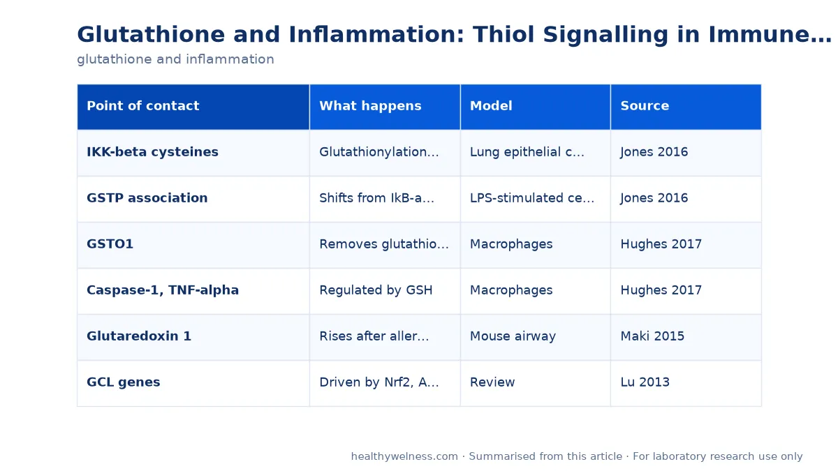
Where glutathione touches the inflammatory machinery
| Point of contact | What happens | Model | Source |
|---|---|---|---|
| IKK-beta cysteines | Glutathionylation, NF-kB restrained | Lung epithelial cells | Jones 2016 |
| GSTP association | Shifts from IkB-alpha to IKK-beta | LPS-stimulated cells | Jones 2016 |
| GSTO1 | Removes glutathione | Macrophages | Hughes 2017 |
| Caspase-1, TNF-alpha | Regulated by GSH | Macrophages | Hughes 2017 |
| Glutaredoxin 1 | Rises after allergen | Mouse airway | Maki 2015 |
| GCL genes | Driven by Nrf2, AP-1, NF-kB | Review | Lu 2013 |
The table is a map rather than a mechanism. Each row is a separate observation in a separate system, and the field has not joined them into one pathway. What they share is the chemistry: a thiol group on glutathione that can form a disulfide with a cysteine on a protein, and a set of enzymes that make and break that bond. Our glutathione research compound is the reduced tripeptide used as a reagent in experiments of this kind.
Certificates are available for listed products — contact us for the latest report.
Why the antioxidant story is incomplete
The simplest account of glutathione and inflammation runs like this: inflammation produces reactive oxygen species, glutathione neutralises them, and so more glutathione means less inflammation. Ghezzi’s review of the lung literature sets out to dismantle that account. It notes that most reviews in the area are biased by a picture in which free radicals cause disease and antioxidants prevent it, and that the role of thiols in immunity requires accepting a more complex picture (Ghezzi, 2011). Reactive oxygen species are part of host defence; removing them indiscriminately would not be expected to be harmless.
Morris and colleagues make the same point at the level of individual immune cells. Their review describes how macrophages, dendritic cells, neutrophils, T cells, B cells and natural killer cells all depend on intracellular and extracellular levels of reactive oxygen and nitrogen species, and how these are buffered by the glutathione and thioredoxin systems together with Nrf2 (Morris et al., 2022). In that framing, glutathione is one of several redox buffers that set the conditions under which immune cells polarise, phagocytose and release cytokines. It is a variable that shapes the response rather than a switch that turns it off.
Measurement makes this harder
Part of the difficulty is that “glutathione level” is not a single quantity. There is reduced glutathione (GSH), its disulfide (GSSG), and glutathione bound to proteins. The mouse airway study measured total glutathione, oxidised glutathione and protein S-glutathionylation separately and found that they moved on different timescales (Maki et al., 2015). A study reporting only total GSH would have missed most of what happened.
S-glutathionylation: the signalling route
S-glutathionylation is the conjugation of glutathione to a reactive cysteine on a protein, forming a mixed disulfide. It protects the cysteine from irreversible oxidation, but it also changes how the protein works. Because it can be reversed by glutaredoxin 1 and related enzymes, it behaves like a regulatory modification, comparable in principle to phosphorylation.
The IKK-beta experiments
NF-kappaB is the transcription factor family most associated with pro-inflammatory gene expression, and its activation depends on inhibitory kappa B kinase beta (IKK-beta). Glutathionylation of IKK-beta inhibits its activity. Jones and colleagues asked whether glutathione S-transferase pi (GSTP), an enzyme that can catalyse glutathionylation under oxidative stress, controls this step (Jones et al., 2016).
- In unstimulated lung epithelial cells, GSTP was associated with IkB-alpha, the inhibitor of NF-kappaB.
- After LPS exposure, that association was rapidly lost and GSTP instead associated with IKK-beta for a prolonged period, while IKK-beta glutathionylation increased.
- Knocking GSTP down with siRNA lowered IKK-beta glutathionylation and increased NF-kappaB nuclear translocation, transcriptional activity and cytokine output.
- A selective GSTP inhibitor, TLK117, also increased LPS-induced NF-kappaB activity.
One finding complicates the story. Both wild-type GSTP and a catalytically inactive Y7F mutant reduced GM-CSF production, which the authors read as evidence that GSTP’s influence involves protein interactions as well as catalysis (Jones et al., 2016). The result is a more interesting model than “glutathione suppresses NF-kappaB”: an enzyme that changes partners when the cell is stimulated, and that acts partly as a scaffold.
Macrophages and GSTO1
Hughes, McGettrick and O’Neill reviewed glutathionylation in macrophages, where activation through Toll-like receptor 4 or phagocytosis generates reactive oxygen species. They describe glutathione as having both positive and negative effects on macrophage proteins: it regulates TNF-alpha secretion, HIF-1-alpha stability, STAT3 phosphorylation and caspase-1 activation, and in Listeria-infected macrophages it modifies the bacterial virulence protein PrfA (Hughes et al., 2017). They also highlight glutathione transferase omega 1 (GSTO1), which removes glutathione from proteins and has been linked to macrophage metabolism, NF-kappaB activation and cell survival.
The macrophage literature thus contains enzymes on both sides of the reaction. GSTP adds glutathione; GSTO1 and glutaredoxin 1 take it off. Which effect dominates depends on which proteins are modified, in which cell, at which moment.
Timing in an airway model
The clearest time-course data come from Maki and colleagues, who sensitised BALB/c mice to ovalbumin, gave three aerosol challenges and then sampled lungs and lavage fluid at 6, 24, 48 and 72 hours and at 8 days, with five mice per group (Maki et al., 2015).
| Time after last challenge | Protein S-glutathionylation | Glutaredoxin 1 |
|---|---|---|
| 6 h | Raised in lung | Raised in lavage fluid and alveolar macrophages |
| 24 h | Attenuated | Not reported as raised in tissue |
| 48–72 h | Not reported as raised | Significantly raised in lung tissue |
Glutaredoxin 1 rose alongside Th2 and NF-kappaB-related cytokines and chemokines. The authors suggested that glutaredoxin 1 and protein glutathionylation status may play important roles in the development of allergic airway inflammation. That is a correlation in one model; the study did not manipulate either variable. It does show why sampling time matters: a single measurement at 24 hours would have suggested that glutathionylation was unchanged.
The supply side: inflammation and glutathione synthesis
The relationship also runs the other way. Glutathione is made in the cytosol in two ATP-dependent steps, and the first, catalysed by glutamate cysteine ligase (GCL), is rate-limiting together with cysteine availability. Lu’s review lists the transcription factors that regulate the GCL subunits and glutathione synthetase: Nrf2 through the antioxidant response element, AP-1, and NF-kappaB (Lu, 2013). The same review lists endotoxaemia among conditions in which glutathione synthesis is dysregulated.
So NF-kappaB can be restrained by glutathionylation of IKK-beta and can, in turn, influence the transcription of the enzymes that make glutathione. Any experiment that changes glutathione and then reads out an inflammatory marker is sitting inside a loop, and an observed effect may be partly downstream of a change in synthesis.
Designing an experiment around these findings
- Measure more than one pool. GSH, GSSG and protein-bound glutathione can diverge, as the airway study showed.
- Sample over time. Glutathionylation changes within hours; enzyme levels follow later.
- Separate catalysis from binding. The GSTP Y7F result shows that an enzyme’s effect may not depend on its catalytic site.
- Prepare solutions fresh. Reduced glutathione oxidises to GSSG in solution, so the reagent itself can change the GSH:GSSG ratio of an experiment. See preparing a glutathione solution and the peptide calculator for concentrations.
Related research
The structure and the unusual gamma-glutamyl bond are covered in what is glutathione, and the enzymes that use it in how glutathione works. For the cysteine-donor comparison, see NAC versus glutathione.
Frequently asked questions
Is glutathione simply an anti-inflammatory antioxidant?
No. Reviews of the field warn against that picture. Ghezzi (2011) describes glutathione as both an antioxidant and a signalling molecule in innate immunity, and Hughes et al. (2017) list macrophage functions, such as TNF-alpha release and caspase-1 activation, that glutathione can drive as well as restrain.
What is S-glutathionylation?
S-glutathionylation is the formation of a mixed disulfide between glutathione and a reactive cysteine on a protein. It can change the protein’s activity and is reversed by enzymes such as glutaredoxin 1. In lung epithelial cells, glutathionylation of IKK-beta was linked to restraint of NF-kappaB signalling (Jones et al., 2016).
Which enzymes add or remove glutathione from proteins?
Glutathione S-transferase pi has been shown to catalyse protein S-glutathionylation under oxidative stress (Jones et al., 2016). Glutaredoxin 1 reverses it, and glutathione transferase omega 1 acts as a deglutathionylating enzyme in macrophages (Hughes et al., 2017).
What animal models link glutathione to airway inflammation?
In BALB/c mice sensitised and challenged with ovalbumin, protein S-glutathionylation in the lung rose at the final challenge and fell by 24 hours, while glutaredoxin 1 rose in lung tissue at 48 and 72 hours alongside Th2 and NF-kappaB-related cytokines (Maki et al., 2015).
Does inflammation change how much glutathione a cell makes?
Synthesis is regulated by the rate-limiting enzyme glutamate cysteine ligase, whose genes are controlled by Nrf2, AP-1 and NF-kappaB (Lu, 2013). Inflammatory transcription factors can therefore influence glutathione supply as well as being influenced by it.
For laboratory research use only. The research materials supplied by HealthyWelness are not approved medicines, foods or cosmetics and are not for human or veterinary use. This article summarises published research and is not medical advice.
Sources
- Ghezzi P. Role of glutathione in immunity and inflammation in the lung. Int J Gen Med. 2011. PMID: 21403800
- Morris G, et al. Redox regulation of the immune response. Cell Mol Immunol. 2022. PMID: 36056148
- Jones JT, et al. Glutathione S-transferase pi modulates NF-κB activation and pro-inflammatory responses in lung epithelial cells. Redox Biol. 2016. PMID: 27058114
- Hughes MM, et al. Glutathione and Glutathione Transferase Omega 1 as Key Posttranslational Regulators in Macrophages. Microbiol Spectr. 2017. PMID: 28102119
- Maki K, et al. Temporal changes in glutaredoxin 1 and protein s-glutathionylation in allergic airway inflammation. PLoS One. 2015. PMID: 25874776
- Lu SC. Glutathione synthesis. Biochim Biophys Acta. 2013. PMID: 22995213