NAD+ Structure: Two Nucleotides Joined by a Pyrophosphate
NAD+ is two nucleotides, NMN and AMP, joined through a pyrophosphate bridge. Formula C21H27N7O14P2, the reactive nicotinamide ring, and how enzymes cut it.

Cover illustration: AI-generated editorial artwork. Conceptual only; not an exact molecular structure, actual laboratory result or evidence of clinical effects.
NAD+ (nicotinamide adenine dinucleotide) is two nucleotides, nicotinamide mononucleotide and adenosine monophosphate, joined through a pyrophosphate bridge. Its formula is C21H27N7O14P2 (PubChem CID 5892), and all of its redox chemistry happens on the positively charged nicotinamide ring.
For laboratory research use only. The research materials supplied by HealthyWelness are not approved medicines, foods or cosmetics and are not for human or veterinary use. This article summarises published research and is not medical advice.
- PubChem records NAD+ (nadide) as CID 5892, with molecular formula C21H27N7O14P2, molecular weight 663.4 g/mol and exact mass 663.109.
- NAD+ consists of a nicotinamide riboside and an adenosine, each carrying a phosphate, linked through a pyrophosphate (diphosphate) bond.
- The nicotinamide ring of NAD+ is a pyridinium cation, described in PubChem’s IUPAC name as 3-carbamoylpyridin-1-ium.
- In mitochondria, NMNAT3 can reversibly cleave NAD+ into nicotinamide mononucleotide and ATP (Hoyland et al., 2024).
- The crystal structure of human ALDH1A3 complexed with NAD+ showed two distinct, coupled conformations of the bound cofactor (Moretti et al., 2016).
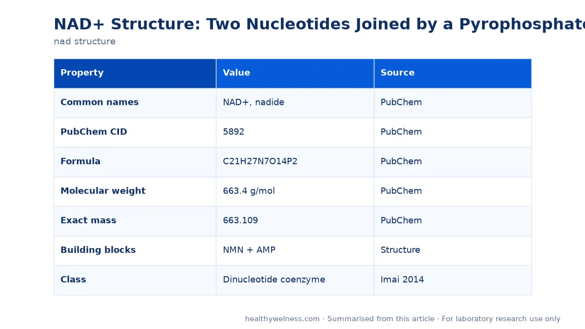
Quick facts
| Property | Value | Source |
|---|---|---|
| Common names | NAD+, nadide | PubChem |
| PubChem CID | 5892 | PubChem |
| Formula | C21H27N7O14P2 | PubChem |
| Molecular weight | 663.4 g/mol | PubChem |
| Exact mass | 663.109 | PubChem |
| Building blocks | NMN + AMP | Structure |
| Class | Dinucleotide coenzyme | Imai 2014 |
These values describe the NAD+ species as PubChem records it. Material supplied as a salt, such as a sodium salt, or as a hydrate has a different formula weight, so the figure for a specific lot belongs on that lot’s documentation rather than in a general table. Our NAD+ research material page lists the product; the certificate gives the form.
Building NAD+ from its parts
It is easiest to read the structure as an assembly of five pieces, from one end to the other.
| Piece | Chemical identity | Role |
|---|---|---|
| Nicotinamide | Pyridine-3-carboxamide | Accepts the hydride |
| Ribose 1 | D-ribose, beta-linked | Carries nicotinamide |
| Pyrophosphate | Two phosphates, P-O-P | Joins the halves |
| Ribose 2 | D-ribose | Carries adenine |
| Adenine | 6-Aminopurine | Binding handle |
Nicotinamide plus the first ribose is nicotinamide riboside; adding a phosphate gives nicotinamide mononucleotide (NMN). Adenine plus the second ribose is adenosine; adding a phosphate gives AMP. Joining the two phosphates releases water and forms the pyrophosphate bond, giving NAD+. PubChem’s IUPAC name spells out all of this: a 6-aminopurin-9-yl group on one oxolane (ribose) ring, a 3-carbamoylpyridin-1-ium group on another, and a methoxy-hydroxyphosphoryl phosphate linking them.
Stereochemistry
Both sugars are D-ribose in the furanose form, and the PubChem name gives each ring the configuration (2R,3S,4R,5R). The glycosidic bond to nicotinamide is the beta anomer, which is the form enzymes recognise. Enzymes that use NAD+ recognise this specific three-dimensional arrangement, so the configuration is part of what defines the active coenzyme rather than a detail of nomenclature.
The reactive end: the nicotinamide ring
NAD+ is named for its oxidised state. The nitrogen of the nicotinamide ring carries a positive charge, making the ring electron-poor and able to accept a hydride (two electrons and a proton) at its C4 position. That converts NAD+ to NADH. The reverse reaction donates the hydride back. Imai and Guarente describe NAD+ as a classical coenzyme mediating many redox reactions in exactly this way (Imai and Guarente, 2014).
The important structural point is that redox chemistry leaves the rest of the molecule intact. The adenosine half and pyrophosphate do not change between NAD+ and NADH, so the same coenzyme molecule can be reduced and re-oxidised many times without being used up. A fuller treatment of that interconversion is in NAD+ and NADH redox.
Where enzymes cut the molecule
The second, non-redox role of NAD+ is as a substrate that is consumed. Here structure matters differently, because each consuming enzyme breaks a specific bond.
| Enzyme | Bond attacked | Source |
|---|---|---|
| Sirtuins | Nicotinamide-ribose | Imai 2014 |
| PARPs | Nicotinamide-ribose | Stromland 2021 |
| CD38, CD157 | Nicotinamide-ribose | Gasparrini 2021 |
| NPP1, CD73 | Phosphate bonds | Gasparrini 2021 |
| NMNAT3 (reverse) | Pyrophosphate | Hoyland 2024 |
Three of the five attack the same bond, the glycosidic link between nicotinamide and its ribose. That bond is high-energy in the sense that breaking it releases enough free energy to drive transfer of the ADP-ribose part onto proteins or water. The release of free nicotinamide from each of these reactions is the reason nicotinamide recycling through the salvage pathway is central to NAD+ maintenance (Stromland et al., 2021).
Gasparrini and colleagues describe the extracellular side: ectoenzymes including CD73, nucleotide pyrophosphatase/phosphodiesterase 1, CD38 and its paralogue CD157 hydrolyse NAD+, generate signalling molecules such as adenosine, and release pyridine metabolites that cells can take up to regenerate NAD+ (Gasparrini et al., 2021). Inside mitochondria, Hoyland and colleagues found that NMNAT3 can run the synthesis reaction backwards, cleaving NAD+ to NMN and ATP and so maintaining what they describe as an additional, virtual NAD+ pool (Hoyland et al., 2024).
How NAD+ sits in a protein
Bound to an enzyme, NAD+ is not a rigid object. In the first crystal structure of human aldehyde dehydrogenase 1A3, solved with NAD+ and the product retinoic acid, the cofactor adopted two different conformations coupled to two positions of the product, which the authors interpreted as snapshots along the catalytic process (Moretti et al., 2016).
This flexibility is a consequence of the structure described above. The pyrophosphate bridge and two ribose rings give the molecule several rotatable bonds, so the nicotinamide end can move relative to the adenosine end while the adenine stays anchored in its pocket. Medicinal chemists have used that architecture as a design template, producing analogues that compete at NAD-dependent enzymes in much the way ATP analogues were developed for kinases (Chen et al., 2008).
Reading a structure diagram correctly
Published diagrams of NAD+ vary in how they draw the charges, and that causes confusion when formulas are compared. Three conventions are common.
| Drawing convention | Net charge shown | What it represents |
|---|---|---|
| Pyridinium plus one phosphate anion | Zero (zwitterion) | The PubChem NAD+ record |
| Pyridinium, both phosphates neutral | Plus one | The “+” in the name |
| Both phosphates ionised | Minus one | Near-neutral pH in solution |
The plus sign in “NAD+” refers to the nicotinamide nitrogen, not to the molecule’s net charge. In water at near-neutral pH the phosphates are ionised, so the molecule as a whole carries a negative charge even though it is still called NAD+. A formula weight copied from one source and a structure copied from another can therefore disagree by the mass of a proton or a counter-ion, which is another reason to take the number from the material’s own documentation.
Why structure matters for handling
- The pyrophosphate bond and the nicotinamide glycosidic bond are the weak points in solution, and both are attacked faster at elevated temperature and away from neutral pH.
- NAD+ absorbs ultraviolet light through its adenine ring, which is how concentration is often checked; NADH has an additional absorbance from its reduced nicotinamide ring, which distinguishes the two forms.
- Biological media containing ectoenzymes will degrade added NAD+, so the starting concentration is not what cells see over time (Gasparrini et al., 2021).
Practical preparation is covered in NAD+ reconstitution, and concentration arithmetic can be done with the peptide calculator by entering the formula weight from the certificate.
Related research
For the compound’s biology see the NAD+ overview. Naming conventions are untangled in NAD versus NAD+, and the phosphorylated relative in NAD+ versus NADPH. The question of whether NAD+ belongs with peptides is answered in is NAD a peptide: it is not, as the structure above makes clear.
Frequently asked questions
What is the chemical structure of NAD+?
NAD+ is a dinucleotide: a nicotinamide mononucleotide and an adenosine monophosphate joined through their phosphate groups into a pyrophosphate bridge. PubChem lists it as CID 5892 with the formula C21H27N7O14P2 and a molecular weight of 663.4 g/mol.
Which part of NAD+ carries the positive charge?
The nitrogen of the nicotinamide ring. The IUPAC name in PubChem describes that ring as 3-carbamoylpyridin-1-ium, the pyridinium cation that gives NAD+ its plus sign and makes it an electron acceptor.
How does NAD+ differ from NADH structurally?
Only in the nicotinamide ring. In reduction the ring accepts a hydride, two electrons and a proton, at its C4 position and loses its aromatic positive charge. The adenosine half and the pyrophosphate bridge are unchanged, which is why the two forms can be interconverted in redox reactions without being consumed (Imai and Guarente, 2014).
Can the two halves of NAD+ be separated by enzymes?
Yes. In mitochondria, NMNAT3 can reversibly cleave NAD+ to nicotinamide mononucleotide and ATP (Hoyland et al., 2024). Outside the cell, ectoenzymes such as CD38, CD73 and NPP1 hydrolyse NAD+ into smaller metabolites (Gasparrini et al., 2021).
Does NAD+ adopt one shape when bound to enzymes?
No. The crystal structure of human ALDH1A3 with NAD+ revealed two different, coupled conformations of the cofactor, which the authors proposed represent two snapshots of the catalytic process (Moretti et al., 2016).
For laboratory research use only. The research materials supplied by HealthyWelness are not approved medicines, foods or cosmetics and are not for human or veterinary use. This article summarises published research and is not medical advice.
Sources
- PubChem. Nadide (NAD+), CID 5892. National Center for Biotechnology Information.
- Imai S, Guarente L. NAD+ and sirtuins in aging and disease. Trends Cell Biol. 2014. PMID: 24786309
- Hoyland LE, et al. Subcellular NAD(+) pools are interconnected and buffered by mitochondrial NAD(). Nat Metab. 2024. PMID: 39702414
- Gasparrini M, et al. Enzymology of extracellular NAD metabolism. Cell Mol Life Sci. 2021. PMID: 33755743
- Moretti A, et al. Crystal structure of human aldehyde dehydrogenase 1A3 complexed with NAD(+) and retinoic acid. Sci Rep. 2016. PMID: 27759097
- Chen L, et al. Nicotinamide adenine dinucleotide based therapeutics. Curr Med Chem. 2008. PMID: 18336280
- Stromland O, et al. The balance between NAD(+) biosynthesis and consumption in ageing. Mech Ageing Dev. 2021. PMID: 34509469