Thymalin vs Thymosin Alpha-1: Extract and Defined Peptide
Thymalin and thymosin alpha-1 compared: a polypeptide thymus extract studied mainly in Russian literature against a single defined 28-residue synthetic peptide.

Cover illustration: AI-generated editorial artwork. Conceptual only; not an exact molecular structure, actual laboratory result or evidence of clinical effects.
Thymalin is a polypeptide complex extracted from thymus tissue, while thymosin alpha-1 is a single defined peptide that can be synthesised to a known sequence. That difference in composition is why one has a characterised mechanism and phase 3 trial data and the other does not.
For laboratory research use only. The research materials supplied by HealthyWelness are not approved medicines, foods or cosmetics and are not for human or veterinary use. This article summarises published research and is not medical advice.
- Thymalin is described as a polypeptide complex isolated from the thymus, with KE and EW named as its active dipeptides (Linkova et al., 2023).
- In the TESTS trial, 28-day all-cause mortality was 23.4% with thymosin alpha-1 and 24.1% with placebo among 1089 analysed participants, a hazard ratio of 0.99 (Wu et al., 2025).
- Thymalin increased CD28 expression in human haematopoietic stem cells 6.8-fold while reducing CD44 and CD117 two- to three-fold (Khavinson et al., 2020).
- Thymosin alpha-1 activated dendritic cells through MyD88-dependent Toll-like receptor signalling and protected transplanted mice from aspergillosis (Romani et al., 2004).
- Thymosin alpha-1’s reported serum half-life is about two hours, with levels returning to baseline within 24 hours (Ancell et al., 2001).
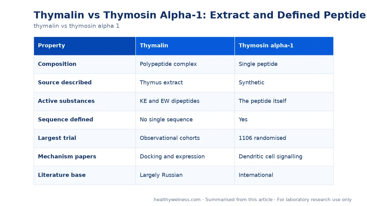
Composition is the dividing line
| Property | Thymalin | Thymosin alpha-1 |
|---|---|---|
| Composition | Polypeptide complex | Single peptide |
| Source described | Thymus extract | Synthetic |
| Active substances | KE and EW dipeptides | The peptide itself |
| Sequence defined | No single sequence | Yes |
| Largest trial | Observational cohorts | 1106 randomised |
| Mechanism papers | Docking and expression | Dendritic cell signalling |
| Literature base | Largely Russian | International |
An extract cannot be characterised the way a defined peptide can. Batch-to-batch composition is not fixed, mass spectrometry returns a mixture, and a mechanism attributed to the preparation may belong to one component, several, or an interaction between them. That is not a criticism of the research so much as a constraint on what any of it can establish.
Material from the thymalin research peptide listing is supplied for laboratory use only. Related profiles are in what is thymosin alpha-1 and the thymosin alpha-1 research review, with the adjacent extract comparison in epithalon vs thymalin and thymalin vs thymulin.
What the thymalin papers report
The modern thymalin literature takes an unusual approach: rather than characterising the extract, it studies two dipeptides identified as its active substances. Linkova and colleagues investigated KE and EW using molecular modelling and docking against double-stranded DNA, reporting GGAG as the preferred binding sequence for EW in classical B-form DNA and GCGC for KE in the curved nucleosomal form. Bioinformatic analysis identified candidate target genes including AKT1 and AKT2, with ACE2 and CYSLTR1 specific to EW and CHUK to KE.
The same paper included a cell experiment, which is the part with direct evidence. Using a lipopolysaccharide-induced inflammation model in human peripheral blood mononuclear cells from four donors, thymalin and both dipeptides reduced synthesis of interleukin-1 beta, interleukin-6 and tumour necrosis factor alpha by 1.4- to 6.0-fold measured by ELISA. The range is wide and the donor count small, but the direction is consistent.
Khavinson and colleagues’ 2020 paper examined human haematopoietic stem cells and reported that thymalin reduced the stem cell marker CD44 and the intermediate differentiation marker CD117 by two- to three-fold while increasing CD28, a marker of mature T lymphocytes, 6.8-fold. The authors state explicitly that this indirectly indicates stimulated differentiation — marker expression rather than a demonstrated lineage transition.
Older clinical reports exist but are harder to assess. A 2002 Russian-language paper reports geroprotective effects of thymalin and epithalamin assessed over six to eight years in 266 elderly people, with reduced acute respiratory disease incidence and lower mortality against a control group. It is an observational report in a non-indexed-abstract format, and the effect sizes claimed are large relative to the design.
What the thymosin alpha-1 record shows
Thymosin alpha-1 has the kind of evidence base an extract cannot generate. Dominari and colleagues’ review describes it as a peptide naturally occurring in the thymus, long recognised for modifying and restoring immune function, and used in immunocompromised states, as a vaccine enhancer, and in sepsis and infections. Ancell and colleagues’ earlier pharmacology review adds the pharmacokinetic detail: rapid absorption, peak serum concentrations within two hours, return to baseline within 24 hours, and a serum half-life of approximately two hours.
The mechanism work is specific. Romani and colleagues showed that thymosin alpha-1 induced functional maturation and interleukin-12 production by fungus-pulsed dendritic cells through the p38 MAPK and NF-kappaB pathway, signalling via MyD88 and involving distinct Toll-like receptors. In vivo, the peptide activated Th1-dependent antifungal immunity and protected highly susceptible mice that had received haematopoietic transplants from aspergillosis. A later review by the same group extended the picture to plasmacytoid dendritic cells via TLR9 and to indoleamine 2,3-dioxygenase induction.
The largest clinical test was negative. TESTS randomised 1106 adults with sepsis across 22 centres in China between 2016 and 2020, with 28-day all-cause mortality as the primary outcome. Mortality was 23.4% with thymosin alpha-1 and 24.1% with placebo, hazard ratio 0.99, p = 0.93, and no secondary or safety outcome differed significantly. Prespecified subgroup analyses suggested differential effects by age and by diabetes status, with interaction p-values of 0.01 and 0.04 — signals the authors present as exploratory rather than conclusions.
| Study | Design | Result |
|---|---|---|
| TESTS 2025 | Phase 3, n=1106 | No mortality difference |
| Romani 2004 | Mouse, cells | TLR-dependent Th1 activation |
| Khavinson 2020 | Human HSC in vitro | CD28 up 6.8-fold |
| Linkova 2023 | PBMC inflammation | Cytokines down 1.4-6x |
Placing the negative phase 3 result next to the positive mechanistic work is the honest reading of this field. A described pathway does not guarantee a clinical outcome, and in this case a well-powered trial did not find one.
What this means for laboratory use
The two materials demand different quality expectations. For a defined synthetic peptide, identity and purity are answerable questions: a single expected mass, an HPLC profile, a stated net peptide content. For a tissue-derived complex, those questions have no single answer, so the relevant controls are different — batch identity, consistency between lots, and documentation of what the preparation contains.
That has a direct consequence for experimental design. A study using a defined peptide can attribute an effect to that molecule. A study using an extract can attribute an effect only to the preparation, which is why the newer thymalin papers work with isolated KE and EW dipeptides instead: a dipeptide is defined, and conclusions about it transfer. Anyone planning work with the extract should consider whether the defined components answer the question better.
Both are supplied lyophilised and both should be reconstituted with gentle swirling and kept cold, with concentrations calculated from the net content on the certificate using the peptide calculator. For the defined peptide, step-by-step handling is in thymosin alpha-1 reconstitution, and general storage practice in lyophilised peptide storage. Certificates are available for listed products — contact us for the latest report.
How to read claims about thymus peptides
Three habits make this literature easier to assess. Check whether a reported effect belongs to the extract or to a defined component, since the newer thymalin papers study KE and EW dipeptides specifically and conclusions about those do not automatically extend to the whole preparation. Check whether a clinical claim comes from a randomised trial or an observational cohort, because the older geroprotective reports are the latter and their effect sizes are large relative to that design. And check the publication language and indexing, since several foundational thymalin reports are Russian-language papers whose methods sections are not available in translation.
Applying the same habits to thymosin alpha-1 gives a different but equally clear picture: a defined sequence, a described signalling pathway with named receptors, a large randomised trial, and a null result on mortality in sepsis. That combination is more informative than a longer list of positive reports, because it shows where the mechanism did and did not translate.
Frequently asked questions
Is thymalin a single peptide?
No. Thymalin is described as a polypeptide complex isolated from the thymus, with KE and EW dipeptides named as its active substances (Khavinson et al., 2020; Linkova et al., 2023). Thymosin alpha-1 is a single defined peptide that can be made synthetically (Dominari et al., 2020).
Which has larger randomised trial data?
Thymosin alpha-1. The TESTS trial randomised 1106 adults with sepsis to thymosin alpha-1 or placebo and found no clear evidence of reduced 28-day mortality, with 23.4% versus 24.1% (Wu et al., 2025). Thymalin’s clinical reports come mainly from Russian-language gerontology literature.
What did the thymalin stem cell study report?
Khavinson and colleagues reported that thymalin reduced expression of CD44 and CD117 in human haematopoietic stem cells by two- to three-fold and increased CD28 expression 6.8-fold, which they interpreted as indirect evidence of stimulated differentiation towards mature T lymphocytes.
Is thymosin alpha-1's mechanism established?
A specific pathway has been described. Romani and colleagues showed that thymosin alpha-1 induced maturation and interleukin-12 production in fungus-pulsed dendritic cells through a p38 MAPK and NF-kappaB pathway, signalling via MyD88 and distinct Toll-like receptors (Romani et al., 2004).
How long does thymosin alpha-1 persist in serum?
The reported serum half-life is approximately two hours, with peak serum concentrations within two hours and a return to baseline within 24 hours (Ancell et al., 2001).
For laboratory research use only. The research materials supplied by HealthyWelness are not approved medicines, foods or cosmetics and are not for human or veterinary use. This article summarises published research and is not medical advice.
Sources
- Khavinson VK, et al. Thymalin: Activation of Differentiation of Human Hematopoietic Stem Cells. Bull Exp Biol Med. 2020. PMID: 33237528
- Linkova N, et al. The Influence of KE and EW Dipeptides in the Composition of the Thymalin Drug on Gene Expression and Protein Synthesis Involved in the Pathogenesis of COVID-19. Int J Mol Sci. 2023. PMID: 37686182
- Dominari A, et al. Thymosin alpha 1: A comprehensive review of the literature. World J Virol. 2020. PMID: 33362999
- Wu J, et al. The efficacy and safety of thymosin alpha1 for sepsis (TESTS): multicentre, double blinded, randomised, placebo controlled, phase 3 trial. BMJ. 2025. PMID: 39814420
- Romani L, et al. Thymosin alpha 1 activates dendritic cells for antifungal Th1 resistance through toll-like receptor signaling. Blood. 2004. PMID: 14982877
- Ancell CD, et al. Thymosin alpha-1. Am J Health Syst Pharm. 2001. PMID: 11381492